 Products Home / Imaging Systems / OCT Imaging Systems & Components / Application Articles / Swept-Source OCT Images of Micro-Retroreflectors
Swept-Source OCT Images of Micro-Retroreflectors

Please Wait
Swept-Source OCT Images of Micro-Retroreflectors in Biological Tissue
Featured Researchers:
Steven N. Ivers, Stephen A. Baranov, Tim Sherlock, Katerina Kourentzi, Paul Ruchhoeft, Richard Willson, and Kirill V. Larin
|
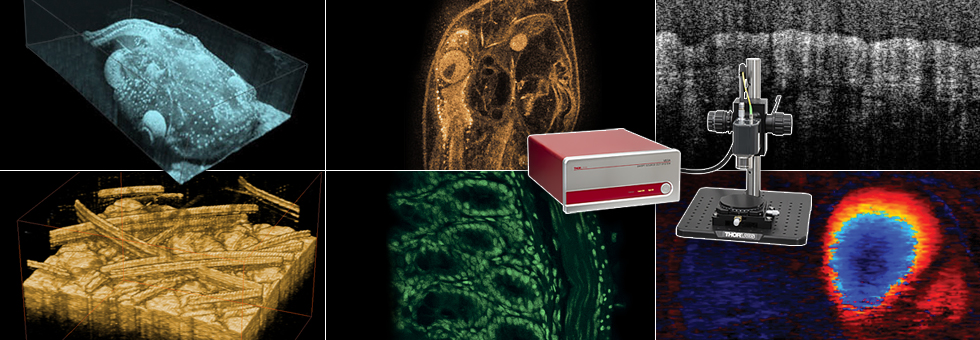
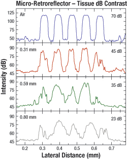
Figure 1.1 Intensity profiles of the micro- Optical Coherence Tomography (OCT) is a high resolution imaging modality that enables nondestructive imaging of a wide range of samples. Recently, a collaborative group of researchers from the University of Houston used a modified Thorlabs high-speed Swept Source OCT (SS-OCT) System (the former OCS1300SS) centered at 1325 nm to image micron-sized retroreflectors through biological tissue. Light reflected by these micro-retroreflectors could yield information on their surrounding environment, forming the basis of a biosensor that provides an alternative method for monitoring the concentration of analytes in the blood such as glucose. Linear, gold, micro-retroreflector arrays were fabricated (5 μm tall, 10 μm wide, and 50 μm long) on silicon wafers and SEM images were taken, as shown in Figure 1.2. Imaging of the entire micro-retroreflector array as a function of depth in porcine skin samples was then carried out using the SS-OCT system to investigate the systems' capability of imaging through highly scattering and turbulent media. To obtain these images, small skin samples (0.5 cm x 0.5 cm) with thicknesses ranging from 0.29 mm to 0.91 mm were excised from a porcine ear, placed on top of the micro-retroreflectors, and kept hydrated in a saline bath during SS-OCT imaging. Figure 1.1 shows 1D intensity profiles of the micro-retroreflectors in air, as well as beneath varying thicknesses of porcine tissue. Each profile was constructed by averaging the intensity data from multiple scans of a given micro-retroreflector row of Figure 1.2. In this study, the minimum level of contrast detected was 20 ± 6.4 dB for porcine tissue. Below this contrast level, the micro-retroreflector structures became obscured due to tissue inhomogeneities. This minimum level of contrast corresponds to a maximum penetration depth of 1.15 mm. By coating the micro-retroreflector arrays with chemical compounds that are selectively sensitive to certain analytes, it may be possible to use them in conjunction with SS-OCT to monitor glucose levels in highly light scattering subcutaneous environments by quantifying reflectivity of the array. This would provide an alternative method to skin puncture for determining blood glucose levels. 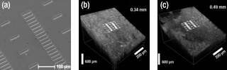
Figure 1.2 SEM images of the linear, gold, micro-retroreflectors used in this study. They were grown on a silicon substrate. These are the 3D SS-OCT images (1 mm x 1 mm x 3 mm) of the micro-retroreflectors, which were placed beneath 0.34 mm and 0.49 mm of porcine tissue, respectively.
|
Reference:
S. N. Ivers et al., Biomedical Optics Express. 1, 367-377 (2010).
| Posted Comments: | |
| No Comments Posted |