Parametric Tissue Imaging


Please Wait
OCT Used for Parametric Imaging of Malignant Tissue
Featured Researchers:
R. A. McLaughlin, L. Scolaro, P. Robbins, C. Saunders, S. L. Jacques, D. D. Sampson
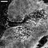
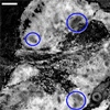
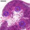
The inability of current clinical imaging techniques to clearly differentiate between healthy and cancerous tissue greatly limits the effectiveness of cancer treatments. During the surgical excision of malignant lymph nodes, which are known to be a common route for the spread of cancer, it is common for patients to also have healthy lymph nodes removed unnecessarily. Removal of lymph nodes can lead to lymphedema, a chronic condition characterized by fluid retention and tissue swelling. Recently, a group of collaborative researchers used a modified Thorlabs Swept-Source Optical Coherence Tomography (SS-OCT) system (central wavelength: 1325 nm, spectral bandwidth: 100 nm) to produce parametric OCT images of human lymph nodes containing malignant cells. These OCT images, which exhibited improved tissue contrast over en face OCT images, potentially hold the key for valuable advancement in cancer treatment. During the study, non-fixed, malignant human axillary lymph nodes were excised from two breast cancer patients. Immediately after excision, the samples were dissected into 2 mm slices, mounted on top of a glass slide, and immersed in glycerol to reduce the refractive index mismatch between the glass slide and tissue sample. The OCT probe was placed underneath the tissue and glass slide, with the probe beam directed upwards. An automated image processing algorithm was used to identify the start of the tissue in each A-scan, and the signal was then analyzed to quantify the rate of OCT signal attenuation over the first 0.5 mm of tissue depth. Cancerous tissue was found to attenuate the OCT signal more rapidly than healthy tissue. The calculated attenuation coefficients were visualized as a parametric image, where the pixel intensity at each (x, y) location indicated the rate of A-scan signal attenuation at that position. Areas In Figure 1, the resulting parametric en face OCT image of a lymph node (frame b) is compared to an H&E histology (frame a) and an en face OCT image (frame c) to assess the technique's ability to differentiate healthy and cancerous tissue. Each lymph node displays areas of malignancy as well as healthy tissue, with several areas of healthy tissue being circled in Figs. 1a and 1b. The parametric OCT image clearly shows a higher level of contrast between healthy and cancerous tissue compared to the en face OCT image. The results shown here demonstrate the ability of parametric OCT to provide a more accurate determination of a cancer patient's condition than en face OCT. This technique could significantly benefit the study, understanding, and treatment of diseases such as cancer.  (c)  (b)  (a) Figure 1. (a) H & E histology, (b) parametric OCT image, and (c) en face OCT image of a malignant human axillary lymph node. The circled areas indicate healthy tissue, which exhibits a lower attenuation coefficient than malignant tissue. Scale bar = 1 mm. |
Reference:
R. A. McLaughlin, L. Scolaro, P. Robbins, C. Saunders, S. L. Jacques, D. D. Sampson, J. Biomed Opt. 15, 046029 (2010).
| Posted Comments: | |
| No Comments Posted |
 Products Home
 OCT Images Malignant Tissue