 Products Home
OCT Application Highlights

Please Wait
Angiography
Art Conservation
Biofilm Imaging
Biomedical OCT
Dentistry
Developmental Biology
Elastography
Industrial OCT
Cochlear Vibrometry
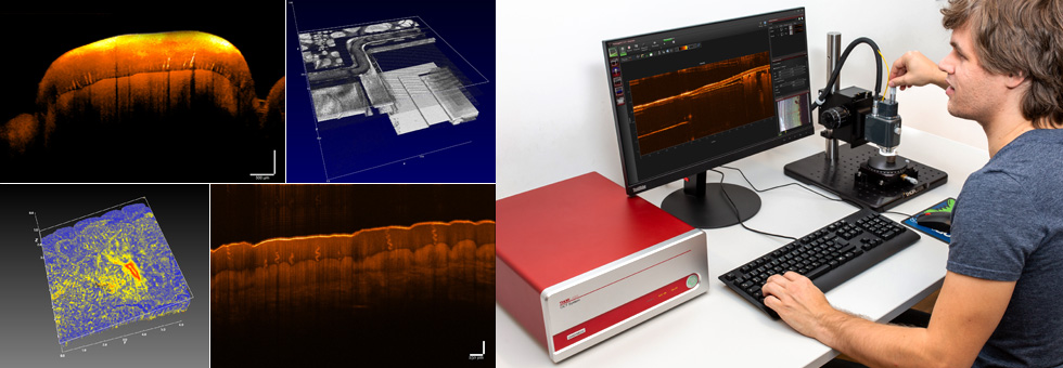
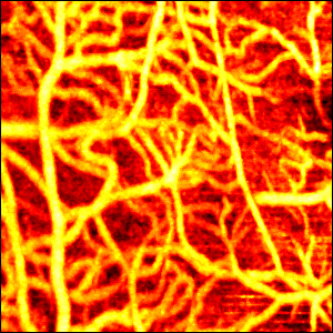
Application Summary
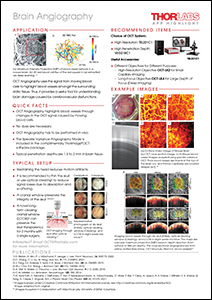
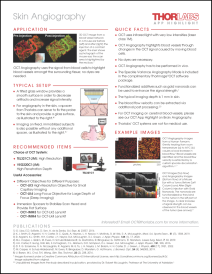
OCT Angiography is used to visualize high-resolution volumetric microvasculature of living organs. The technique uses backscattered light from blood cells to highlight blood vessels from the surrounding tissue; no dyes are needed. Further advantages are the noninvasive visualization of capillary perfusion in vivo. For more information, please view the ![]() Brain Angiography and
Brain Angiography and ![]() Skin Angiography App Highlights.
Skin Angiography App Highlights.
Publications
Akif A, Walek K, Polucha C, Lee J. "Doppler OCT clutter rejection using variance minimization and offset extrapolation." Biomedical Optics Express. 2018 November 1; 9(11): 5340-5352
Liba O, Lew MD, SoRelle ED, Dutta R, Sen D, Moshfeghi DM, Chu S, de la Zerda A. "Speckle-modulating optical coherence tomography in living mice and humans." Nature Communications. 2017 June 20; 8: 15845.
Tang J, Erdener SE, Sunil S, Boas DA. "Normalized field autocorrelation function-based optical coherence tomography three-dimensional angiography." Journal of Biomedical Optics. 2019 March 13; 24(3): 036005.
Dutta R, Liba O, SoRelle ED, Winetraub Y, Ramani V, Jeffrey SS, Sledge GW, de la Zerda A. "Real-time detection of circulating tumor cells in living animals using functionalized large gold nanorods." bioRxiv. 2018 December 17; 498188.
Srinivasan VJ, Jiang JY, Yaseen MA, Radhakrishnan H, Wu W, Barry S, Cable AE, and Boas DA. "Rapid volumetric angiography of cortical microvasculature with optical coherence tomography." Optics Letters. 2010 January 1; 35(1): 43-45.
Collins HA, Khurana M, Moriyama EH, Mariampillai A, Dahlstedt E, Balaz M, Kuimova MK, Drobizhev M, Yang VXD, Phillips D, Rebane A, Wilson BC, Anderson HL. "Blood-vessel closure using photosensitizers engineered for two-photon excitation." Nature Photon. 2008 May 30; 2: 420-424.
Mariampillai A, Standish BA, Moriyama EH, Khurana M, Munce NR, Leung MKK, Jiang J, Cable A, Wilson BC, Vitkin IA, and Yang VXD. "Speckle variance detection of microvasculature using swept-source optical coherence tomography." Optics Letters. 2008 July 1; 33(13): 1530-1532.
Aigner M, Salaberger D, Buchsbaum A, Heise B, Schausberger SE, Köpplmayr T, Lang C, Leitner M, Stifter D, Burzic I, Miethlinger J. "The influence of glass fibers on elongational viscosity studied by means of optical coherence tomography and X-ray computed tomography." AIP Conference Proceedings. 2014; 1593: 217.
Casper M, Schulz-Hildebrandt H, Evers M, Birngruber R, Manstein D, Hüttmann G. "Optimization-based vessel segmentation pipeline for robust quantification of capillary networks in skin with optical coherence tomography angiography." Journal of Biomedical Optics. 2019 April 30; 24(4): 046005.
Casper MJ, Glahn J, Evers M, Schulz-Hildebrandt H, Kositratna G, Birngruber R, Hüttmann G, Manstein D. "Capillary Refill—The Key to Assessing Dermal Capillary Capacity and Pathology in Optical Coherence Tomography Angiography." Lasers in Surgery and Medicine. 2019 November 21; 52: 653-658.
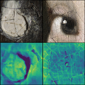
Application Summary
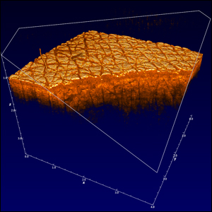
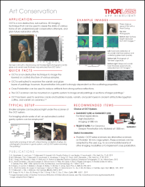
OCT is a non-destructive imaging technique that acquires 3D sub surface information. Within the field of art conservation OCT has a unique significance for that reason as sublayers would remain invisible without the cross-sectional information. OCT assists to assess the state of various forms of art e.g. for paintings, understand past conservation attempts, and plan further restoration efforts. For more information, please view the ![]() Art Conservation App Highlight.
Art Conservation App Highlight.
Publications
Koch-Dandolo CL, Lopez M, Fukunaga K, Ueno Y, Pillay R, Giovannacci D, Du YL, Bai X, Menu M, Detalle V. "Toward a multimodal fusion of layered cultural object images: Complementarity of optical coherence tomography and terahertz time-domain imaging in the heritage field." Applied Optics. 2019 February 10; 58(5); 1281-1290.
Adler DC, Stenger J, Gorczynska I, Lie H, Hensick T, Spronk R, Wolohojian S, Khandekar N, Jiang JY, Barry S, Cable AE, Huber R, Fujimoto JG. "Comparison of three-dimensional optical coherence tomography and high resolution photography for art conservation studies." Optics Express. 2007 November 26; 15(24): 15972-15986.
Callewaert T, Dik J, Kalkman J. "Segmentation of thin corrugated layers in high-resolution OCT images." Optics Express. 2017 December 25; 25(26): 32816-32828.
Vandivere A, van Loon A, Callewaert T, Haswell R, Gaibor ANP, van Keulen H, Leonhardt E, Dik J. "Fading into the background: the dark space surrounding Vermeer’s Girl with a Pearl Earring." Heritage Science. 2019 September 16; 7: 69.
Elkhuizen WS, Callewaert TWJ, Leonhardt E, Vandivere A, Song Y, Pont SC, Geraedts JMP, Dik J. "Comparison of three 3D scanning techniques for paintings, as applied to Vermeer’s ‘Girl with a Pearl Earring’." Heritage Science. 2019 November 4; 7: 89.
Dal Fovo A, Tserevelakis GJ, Papanikolaou A, Zacharakis G, Fontana R. "Combined photoacoustic imaging to delineate the internal structure of paintings." Optics Letters. 2019 February 15; 44(4): 919-922.
Callewaert T, Guo J, Harteveld G, Vandivere A, Eisemann E, Dik J, Kalkman J. "Multi-scale optical coherence tomography imaging and visualization of Vermeer’s Girl with a Pearl Earring." Optics Express. 2020 August 31; 28(18): 26239-26256.
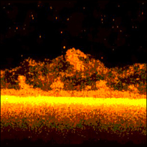
Application Summary
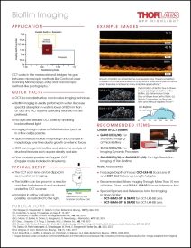
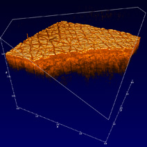
Biofilms form in water systems and are typically an unwanted contamination. There is an increasing interest in understanding the formation and the removal of such biofilms from water systems. For monitoring the morphology of biofilms, OCT has gained much interest in recent years. Here, OCT serves as a complementary imaging method to established methods such as confocal and widefield microscopy and opens up the possibility to monitor the morphology of biofilms on large scales as well as their dynamics in real time. For more information, please view the ![]() Biofilm Application Notes or
Biofilm Application Notes or ![]() Biofilm App Highlight.
Biofilm App Highlight.
Publications
F. Blauert, H. Horn, M. Wagner. "Time-resolved biofilm deformation measurements using optical coherence tomography." Biotechnology and Bioengineering. 2015 March 18; 112 (9): 1893-1905.
M. Wagner, D. Taherzadeh, C. Haisch, H. Horn. "Investigation of the mesoscale structure and volumetric features of biofilms using optical coherence tomography." Biotechnology and Bioengineering. 2010 Aug 17; 107 (5): 844-853.
K.J. Martin, D. Bolster, N. Derlon, E. Morgenroth, R. Nerenberg. "Effect of fouling layer spatial distribution on permeate flux: A theoretical and experimental study." Journal of Membrane Science. 2014 Dec 1; 471 130-137.
L. Fortunato, A. Qamar, Y. Wang, S. Jeong, T. Leiknes "In-situ assessment of biofilm formation in submerged membrane system using optical coherence tomography and computational fluid dynamics." Journal of Membrane Science. 2017 Dec 1; 521: 84-94.
C. Picioreanu, F. Blauert, H, Horn, M. Wagner. "Determination of mechanical properties of biofilms by modelling the deformation measured using optical coherence tomography." Water Research. 2018 Nov 15; 145: 588-598.
M.C. Leite De Andrade; et al. "A new approach by optical coherence tomography for elucidating biofilm formation by emergent Candida species." PLOS ONE. 2017 Nov 16; 12 (11): e0188020.
Application Summary
Due to the versatility of OCT, many researchers and professionals in biological and medical fields employ it in order to image a wide range of tissue types in a non-destructive and contactless way. While the technology is still in the field of theoretical or experimental research in some areas, its capabilities have already been proven in others.
Biomedical OCT areas of interest include:
- Cochlea
- Organ Tissue
- Skin
- Nervous System
- Microvasculature
Publications
Zhang W, Li Y, Nguyen VP, Huang Z, Liu Z, Wang X, Paulus YM. "High-resolution, in vivo multimodal photoacoustic microscopy, optical coherence tomography, and fluorescence microscopy imaging of rabbit retinal neovascularization." Nature. 2018 December 5; 7: 103.
Nguyen VP, Li Y, Zhang W, Wang X, Paulus YM. "Multi-wavelength, en-face photoacoustic microscopy and optical coherence tomography imaging for early and selective detection of laser induced retinal vein occlusion." Biomedical Optics Express. 2018 December 1; 9(12): 5915-5938.
Capowski EE, Samimi K, Mayerl SJ, Phillips MJ, Pinilla I, Howden SE, Saha J, Jansen AD, Edwards KL, Jager LD, Barlow K, Valiauga R, Erlichman Z, Hagstrom A, Sinha D, Sluch VM, Chamling X, Zack DJ, Skala MC, Gamm DM. "Reproducibility and staging of 3D human retinal organoids across multiple pluripotent stem cell lines." Development. 2019 January 9; 146: dev171686.
Leite de Andrade MC, Soares de Oliveira MA, Graciano dos Santos FA, Vilela PBX, da Silva MN, Macêdo DPC, Neto RGL, Neves HJP, Brandão ISL, Chaves GM, de Araujo RE, Neves RP. "A new approach by optical coherence tomography for elucidating biofilm formation by emergent Candida species." Plos One. 2017 November 16; 12(11): e-188020.
Kerse C, Kalaycioglu H, Elahi P, Çetin B, Kesim DK, Akçaalan O, Yavas S, Asik MD, Öktem B, Hoogland H, Holzwarth R, Ilday FO. "Ablation-cooled material removal with ultrafast bursts of pulses." Nature. 2016 July 13; 537: 84-88.
Ahsen OO, Tao YK, Potsaid BM, Sheikine Y, Jiang J, Grulkowski I, Tsai T, Jayaraman V, Kraus MF, Connolly JL, Hornegger J, Cable A, Fujimoto JG. "Swept source optical coherence microscopy using a 1310 nm VCSEL light source." Optics Express. 2013 July 29; 21(15) 18021-18033.
Potsaid B; Jayaraman V; Fujimoto JG; Jiang J; Heim PJS; Cable AE. "MEMS tunable VCSEL light source for ultrahigh speed 60kHz - 1MHz axial scan rate and long range centimeter class OCT imaging." SPIE Proceedings. 2012 February 8; 8213.
Tsai T, Tao YK, Potsaid BM, Jayaraman V, Kraus MF, Heim PJS, Hornegger J, Mashimo H, Cable AE, Fujimoto JG. "Ultrahigh speed endoscopic optical coherence tomography using micro-motor imaging catheter and VCSEL technology." SPIE Proceedings. 2013 March 20; 8571.
Jayaraman V, Potsaid B, Jiang J, Cole GD, Robertson ME, Burgner CB, John DD, Grulkowski I, Choi W, Tsai TH, Liu J, Stein BA, Sanders ST, Fujimoto JG, Cable AE. "High-speed ultra-broad tuning MEMS-VCSELs for imaging and spectroscopy." SPIE Proceedings. 2013 May 17; 8763.
Tsai T, Potsaid B, Tao YK, Jayaraman V, Jiang J, Heim PJS, Kraus MF, Zhou C, Hornegger J, Mashimo H, Cable AE, Fujimoto JG. "Ultrahigh speed endoscopic optical coherence tomography using micromotor imaging catheter and VCSEL technology." Biomedical Optics Express. 2013 July 1; 4(7): 1119-1132.
Choi W, Potsaid B, Jayaraman V, Baumann B, Grulkowski I, Liu JJ, Lu CD, Cable AE, Huang D, Duker JS, Fujimoto JG. "Phase-sensitive swept-source optical coherence tomography imaging of the human retina with a vertical cavity surface-emitting laser light source." Optics Letters. 2013 February 1; 38(3): 338-340.
Li Q, Onozato ML, Andrews PM, Chen C, Paek A, Naphas R, Yuan S, Jiang J, Cable A, Chen Y. "Automated quantification of microstructural dimensions of the human kidney using optical coherence tomography (OCT)." Optics Express. 2009 August 31; 17(18): 16000-16016.
Chen Y, Aguirre AD, Ruvinskaya L, Devor A, Boas DA, Fujimoto JG. "Optical coherence tomography (OCT) reveals depth-resolved dynamics during functional brain activation." Journal of Neuroscience Methods. 2009 March 30; 178(1): 162-173.
Yuan S, Li Q, Jiang J, Cable A, Chen Y. "Three-dimensional coregistered optical coherence tomography and line-scanning fluorescence laminar optical tomography." Optics Letters. 2009 June 1; 34(11): 1615-1617.
Andrews PM, Chen Y, Onozato ML, Huang S, Adler DC, Huber RA, Jiang J, Barry SE, Cable AE, Fujimoto JG. "High-resolution optical coherence tomography imaging of the living kidney." Laboratory Investigation. 2008 February 11; 88: 441-449.
Wen X, Jacques SL, Tuchin VV, Zhu D. "Enhanced optical clearing of skin in vivo and optical coherence tomography in-depth imaging." Journal of Biomedical Optics. 2012 June 13; 17(6): 066022.
Grulkowski I, Liu JJ, Potsaid B, Jayaraman V, Lu CD, Jiang J, Cable AE, Duker JS, Fujimoto JG. "Retinal, anterior segment and full eye imaging using ultrahigh speed swept source OCT with vertical-cavity surface emitting lasers." Biomedical Optics Express. 2012 November 1; 3(11): 2733-2751.
Grulkowski I, Potsaid B, Liu JJ, Jayaraman V, Cable AE, Jiang J, Kraus MF, Hornegger J, Fujimoto JG. "Ophthalmic Applications of Ultrahigh Speed OCT Using VCSEL Light Source Technology." Investigative Ophthalmology & Visual Science. 2012 March; 53(14): 5258.
Grulkowski I, Liu JJ, Zhang JY, Potsaid B, Jayaraman V, Cable AE, Duker JS, Fujimoto JG. "Reproducibility of a Long-Range Swept-Source Optical Coherence Tomography Ocular Biometry System and Comparison with Clinical Biometers." American Academy of Ophthalmology. 2013 June 5; 120(11): 2184-2190.
Lu C, Kraus M, Grulkowski I, Liu J, Potsaid B, Jayaraman V, Cable A, Hornegger J, Duker J, Fujimoto J. "Handheld High Speed 500 kHz Swept Source OCT Device Using a Micro Scanning Mirror." Investigative Ophthalmology & Visual Science. 2013 June; 54(15): 1489.
Potsaid B, Liu J, Choi W, Grulkowski I, Jayaraman V, Jiang J, Heim P, Jay Duker J, Cable A, Fujimoto J. "VCSEL Laser Technology for Ultrahigh Speed and Extended Depth Range OCT Imaging of the Retina and Anterior Eye." Investigative Ophthalmology & Visual Science. 2013 June; 54(15): 1491.
Grulkowski I, Liu J, Potsaid B, Jayaraman V, Cable A, Kraus M, Hornegger J, Duker J, Huang D, Fujimoto J. "Three-Dimensional Biometric Measurements of Accommodation Using Full-Eye-Length Swept-Source OCT." Investigative Ophthalmology & Visual Science. 2013 June; 54(15): 381.
Dhalla A, Liu J, Mohler K, Potsaid B, Lu C, Jayaraman V, Cable A, Huang D, Fujimoto J. "Ultrahigh speed polarization sensitive OCT of the anterior and posterior eye using a 1050 nm VCSEL light source." Investigative Ophthalmology & Visual Science. 2013 June; 54(15): 1492.
Lu CD, Grulkowski I, Liu JJ, Kraus MF, Potsaid B, Jayaraman V, Cable A, Hornegger J, Duker JS, Fujimoto JG. "Handheld High Speed Swept Source Optical Coherence Tomography at 1050nm." 2012 March; 53(14): 2143.
Potsaid B, Gorczynska I, Srinivasan VJ, Chen Y, Jiang J, Cable A, Fujimoto JG. "Ultrahigh speed Spectral / Fourier domain OCT ophthalmic imaging at 70,000 to 312,500 axial scans per second." Optics Express. 2008 September 15; 16(19): 15149-15169.
Srinivasan VJ, Huber R, Gorczynska I, Fujimoto JG, Jiang JY, Reisen P, Cable AE. "High-speed, high-resolution optical coherence tomography retinal imaging with a frequency-swept laser at 850 nm." Optics Letters. 2007 February 15; 32(4): 361-363.
Carolus AE, Lenz M, Hofmann M, Welp H, Schmieder K, Brenke C. "High-resolution in vivo imaging of peripheral nerves using optical coherence tomography: a feasibility study." Journal of Neurosurgery. 2019 April 26; 132(6): 1907-1913.
Argarini R, McLaughlin RA, Joseph SZ, Naylor LH, Carter HH, Yeap BB, Jansen SJ, Green DJ. "Optical coherence tomography: a novel imaging approach to visualize and quantify cutaneous microvascular structure and function in patients with diabetes." BMJ Open Diabetes Research & Care. 2020 August 26; 8: e001479.
Li X, Zhang W, Wang WY, Wu X, Li Y, Tan X, Matera DL, Baker BM, Paulus YM, Fan X, Wang X. "Optical coherence tomography and fluorescence microscopy dual-modality imaging for in vivo single-cell tracking with nanowire lasers." Biomedical Optics Express. 2020 July 1; 11(7): 3659-3672.
Zhang W, Li Y, Yu Y, Derouin K, Qin Y, Nguyen VP, Xiaobo X, Wang X, Paulus YM. "Simultaneous photoacoustic microscopy, spectral-domain optical coherence tomography, and fluorescein microscopy multi-modality retinal imaging." Photoacoustics. 2020 June 6; 20: 100194.
Application Summary
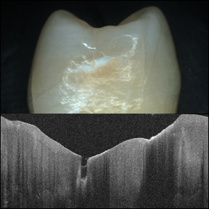
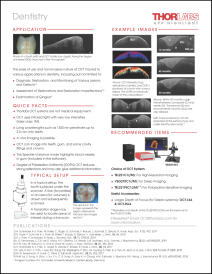
The ease of use and non-invasive nature of OCT has led to various applications in dentistry, including but not limited to:
- The diagnosis, restoration, and monitoring of various lesions and defects;
- The assessment of restorations and restoration imperfections;
- The assessment of restorations and restoration imperfections; and
- The examination of gingiva.
For more information, please view the ![]() Dentistry App Highlight.
Dentistry App Highlight.
Publications
Leiss-Holzinger E, Wiesauer K, Stephani H, Heise B, Stifter D, Kriechbaumer B, Spachinger SJ, Gusenbauer C, Withalm G. "Imaging of the inner structure of cave bear teeth by novel non-destructive techniques." Palaeontologia Electronica. 2015 January 21; 18.1.1T
Seidemannm MR, Haak R, Olms C. "Pilotuntersuchung zur Bewertung von Grenzflächen mittels OCT: Belastung einer Resin-Nano-Keramik auf einteiligen ZrO2-Implantaten." Deutscher Ärzteverlag . 2017; 33 (3): 202-211.
Schneider H, Park K, Hafer M, Ruger C, Schmalz G, Krause F, Schmidt J, Ziebolz D, Haak R. "Dental Applications of Optical Coherence Tomography (OCT) in Cariology." Appl. Sci.. 2017 May 3; 7(5) 472.
Park K, Schneider H, Haak R. "Assessment of interfacial defects at composite restorations by swept source optical coherence tomography." J. of Biomedical Optics. 2013 July 22; 18(7) 076018-1 - 076018-5.
Fernandes LO, Mota CCBO, Oliveira HO, Neves JK, Santiago LM, Gomes ASL. "Optical coherence tomography follow-up of patients treated from periodontal disease." J. Biophotonics. 2019 February; 12(2) e201800209.
Sahyoun CC, Subhash HM, Peru D, Ellwood R, Zaidel L, Kilpatrick L, Pierce MC. "Effect of Optical Coherence Tomography Resolution on the Assessment of Dental Enamel Indications." OSA Technical Digest (Optical Society of America, 2018). 2018 April 3-6;paper JTh3A.5.
S. Lee; et al. "Non-Ionized, High-Resolution Measurement of Internal and Marginal Discrepancies of Dental Prosthesis Using Optical Coherence Tomography." IEEE Access. 2018 December 24; 7 6209-6218.
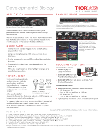
Application Summary
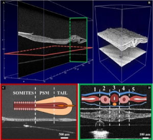
Animal models are studied to understand biological phenomena and transfer the findings to human biology and medicine. The ease of use and non-invasive nature of OCT has made it an indispensable tool that allows researchers to image animal models in-vivo and over the course of the animals’ lives into adulthood. For more information, please view our ![]() Developmental Biology App Highlight.
Developmental Biology App Highlight.
Publications
Wangpraseurt D, Jacques S, Lyndby N, Holm JB, Pages CF, Kühl M. "Microscale light management and inherent optical properties of intact corals studied with optical coherence tomography." Journal of the Royal Society Interface. 2019 Feb 13; 16(151)
Marrese M, Antonovaite N, Nelemans BKA, Smit TH, Iannuzzi D. "Micro-indentation and optical coherence tomography for the mechanical characterization of embryos: Experimental setup and measurements on fixed chicken embryos." Acta Biomaterialia. 2019 Oct 1; 97 (1): 524-534.
Comanns P, Buchberger G, Buchsbaum A, Baumgartner R, Kogler A, Bauer S, Baumgartner W. "Directional, passive liquid transport: the Texas horned lizard as a model for a biomimetic ‘liquid diode’." Journal of the Royal Society Interface. 2015 Aug 6; 12(109).
Dutta R, Liba O, SoRelle ED, Winetraub Y, Ramani VC, Jeffrey SS, Sledge GW, de la Zerda A. "Real-Time Detection of Circulating Tumor Cells in Living Animals Using Functionalized Large Gold Nanorods." Nano Lett.. 2019 March 21; 19 (4): 2334-2342.
Larina IV, Sudheendran N, Mohamad Ghosn, Jiang J, Cable A, Larin KV, Dickinson ME. "Live imaging of blood flow in mammalian embryos using Doppler swept-source optical coherence tomography." J. of Biomedical Optics. 2008 Nov 1; 13(6): 060506.
Mariampillai A, Standish BA, Munce NR, Randall C, Liu G, Jiang JY, Cable AE, Vitkin IA, Yang VXD. " Doppler optical cardiogram gated 2D color flow imaging at 1000 fps and 4D in vivo visualization of embryonic heart at 45 fps on a swept source OCT system." Optics Express. 2007 February 19; 15(4): 1627-1638.
Davis AM, Rothenberg FG, Shepherd N, Izatt JA. "In vivo spectral domain optical coherence tomography volumetric imaging and spectral Doppler velocimetry of early stage embryonic chicken heart development." Journal of the Optical Society of America. 2008 December 1; 25(12): 3134-3143.
Davis A, Izatt JA, Rothenberg F. " Quantitative Measurement of Blood Flow Dynamics in Embryonic Vasculature Using Spectral Doppler Velocimetry." The Anatomical Record. 2009 Feb 26; 292(3): 311-319.
Jenkins SA, Porter TE. " Ontogeny of the hypothalamo–pituitary–adrenocortical axis in the chicken embryo: a review." Domestic Animal Endocrinology. 2004 May; 26(4): 267-275.
K. Courchaine, M.J. Gray, K. Beel, K. Thornburg, S. Rugonyi. " 4-D Computational Modeling of Cardiac Outflow Tract Hemodynamics over Looping Developmental Stages in Chicken Embryos." J. Cardiovasc. Dev. Dis.. 2019 Feb 27; 6 (1): 11.
M.J. Wolf. " 3D imaging of a beating fly heart (upper center) using optical coherence tomography, a technique similar to echocardiography." Duke Today. 2015 February 3.
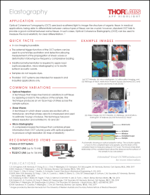
Application Summary
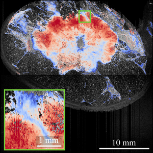
Optical coherence elastography quantifies the elasticity of biological tissue by using Doppler optical coherence tomography (OCT) to measure the local tissue displacement as a function of the applied stress. For more information, please view the ![]() Elastography App Highlight.
Elastography App Highlight.
Publications
Hepburn MS, Wijesinghe P, Chin L, Kennedy BF. "Analysis of spatial resolution in phase-sensitive compression optical coherence elastography." Biomedical Optics Express. 2019; 10(3): 1496-1513.
Kennedy KM, Ford C, Kennedy BF, Bush MB, Sampson DD. "Analysis of mechanical contrast in optical coherence elastography." Journal of Biomedical Optics. 2013 November 12; 18(12): 121508.
Moon S, Chen Z. "Phase-stability optimization of swept-source optical coherence tomography." Biomedical Optics Express. 2018; 9(11): 5280-5295.
Krajancich B, Curatolo A, Fang Q, Zilkens R, Dessauvagie BF, Saunders CM, Kennedy BF. "Handheld optical palpation of turbid tissue with motion-artifact correction." Biomedical Optics Express. 2019; 10(1): 226-241.
Es’haghian S, Kennedy KM, Gong P, Li Q, Chin L, Wijesinghe P, Sampson DD, McLaughlin RA, Kennedy BF. "In vivo volumetric quantitative microelastography of human skin." Biomedical Optics Express. 2017; 8(5): 2458-2471.
Nebelung S, Brill N, Müller F, Tingart M, Pufe T, Merhof D, Schmitt R, Jahr H, Truhn D. "Towards Optical Coherence Tomography-based elastographic evaluation of human cartilage." Journal of the Mechanical Behavior of Biomedical Materials. 2016 March; 56: 106-119.
Düwel D, Otte C, Schulz K, Saatho T, Schlaefer A. "Towards contactless optical coherence elastography with acoustic tissue excitation." Current Directions in Biomedical Engineering. 2015 September 12; 1(1): 215-219.
Allen WM, Kennedy KM, Fang Q, Chin L, Curatolo A, Watts L, Zilkens R, Chin SL, Dessauvagie BF, Latham B, Saunders CM, Kennedy BF. "Wide-field quantitative micro-elastography of human breast tissue." Biomedical Optics Express. 2018; 9(3): 1082-1096.
Es'haghian S, Gong P, Chin L, Harms KA, Murray A, Rea S, Kennedy BF, Wood FM, Sampson DD, McLaughlin RA. "Investigation of optical attenuation imaging using optical coherence tomography for monitoring of scars undergoing fractional laser treatment." Journal of Biophotonics. 2017 April; 10(4): 511-522.
Jonas S, Bhattacharya D, Khokha MK, Choma MA. "Microfluidic characterization of cilia-driven fluid flow using optical coherence tomography-based particle tracking velocimetry." Biomedical Optics Express. 2011; 2(7): 2022-2034.
Kataja M, Haavisto S, Salmela J, Lehto R, Koponen A. "Characterization of micro-fibrillated cellulose fiber suspension flow using multi scale velocity profile measurements." Nordic Pulp & Paper Research Journal. 2018 July 19; 32(3): 473-482.
Meeuw H, Korbelin J, von Bernstorff D, Augustin T, Liebig WV, Fiedler B. "Smart dispersion: Validation of OCT and impedance spectroscopy as solutions for in-situ dispersion analysis of CNP/EP-composites." Materialia. 2018; 1: 185-197.
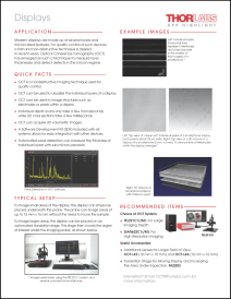
Application Summary
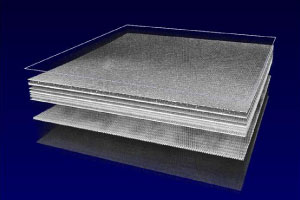
OCT has already proven to be an excellent tool in the industrial field to assess several sample or product characteristics in a fully non-destructive and contactless way. The range of applications spans from film thickness measurements to defect or particle detection and evaluation or control of geometric parameters. OCT can be applied in all stages of the production life cycle, including early R&D, prototyping, and final product evaluation and single-unit quality control. Industrial applications for OCT include but are not limited to:
- Displays
- Pharmaceuticals
- Contact Lens Metrology
- Packaging
- Industrial Paints and Varnish
- Thin Films
- CAD/CAM
- 3D Printing
- Composite Materials
- Aerospace
- Welding and Keyhole Mapping
- Meat Quality Control
For more information, please view our ![]() OCT Displays App Highlight.
OCT Displays App Highlight.
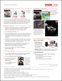
Application Summary
OCT is a powerful technique used to understand the mechanics of hearing, as it is used to locate specific structures within the cochlea via 2D imaging and to provide vibrational data (amplitude and phase shift) at these specific locations. With this information, medical researchers can better analyze disfunction and anomalies of the hearing organ and improve upon treatment methods in the future. For more information, please view the ![]() Vibrometry App Highlight.
Vibrometry App Highlight.
Publications
Cooper NP, Vavakou A, van der Heijden M. "Vibration hotspots reveal longitudinal funneling of sound-evoked motion in the mammalian cochlea." Nature Communications. 2018 August 3; 9: 3054.
Nuttall AL, Ricci AJ, Burwood G, Harte JM, Stenfelt S, Cayé-Thomasen P, Ren T, Ramamoorthy S, Zhang Y, Wilson T, Lunner T, Moore BCJ, Fridberger A. "A mechanoelectrical mechanism for detection of sound envelopes in the hearing organ." Nature Communications. 2018 October 9; 9: 4175.
Ton Y, Sakamoto T, Nakagawa T, Adachi T, Taniguchi M, Torii H, Hamaguchi K, Kitajiri S, Ito J. "In Vivo Imaging of Mouse Cochlea by Optical Coherence Tomography." Otology & Neurology. 2014 February; 35(2): e84-89.
Lin NC, Fallah E, Strimbu CE, Hendon CP, Olson ES. "Scanning optical coherence tomography probe for in vivo imaging and displacement measurements in the cochlea." Biomedical Optics Express. 2019 February 1; 10(2): 1032-1043.
Ling WA, Ellerbee AK. "The effects of reduced bit depth on optical coherence tomography phase data." Optics Express. 2012 July 2; 20(14): 15654-15668.
Pau HW, Lankenau E, Just T, Behrend D, Hüttmann G. "Optical coherence tomography as an orientation guide in cochlear implant surgery?" Acta Oto-Laryngologica. 2009 July 8; 127(9): 907-913.
Vavakou A, Cooper NP, van der Heijden M. "The frequency limit of outer hair cell motility measured in vivo." eLife. 2019 September 24; 8: e47667.
| Posted Comments: | |
| No Comments Posted |